|
 Before proceeding with the presentation of preventive measures that prevent the development of diseases of the gastrointestinal tract, it is necessary to at least briefly dwell on its anatomical and physiological characteristics. Before proceeding with the presentation of preventive measures that prevent the development of diseases of the gastrointestinal tract, it is necessary to at least briefly dwell on its anatomical and physiological characteristics.
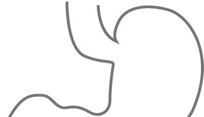
The human stomach is located between the end of the esophagus and the initial part of the duodenum. In it, two surfaces are distinguished - the front and rear, two edges, or two curvatures - small and large, and sections - the entrance part, the bottom (vault), the body and the exit part.
The entrance part is also called cardiac, or heart ("cardia" - in Greek "heart"), since it is closer to the heart. This is where food comes from the esophagus. This is the most basic department.
Then comes the bottom, or vault, - the domed part, located slightly to the left of the entrance.
The exit part, through which food passes into the duodenum, is also called pyloric (“pylorus” - Latin for “gatekeeper”). This is the end of the stomach.
The length of a moderately distended stomach in adults is 22-23 centimeters, the diameter at its widest point is 9-10 centimeters, and the capacity is 3 liters. The capacity varies depending on individual characteristics, as well as the amount of liquid drunk, food eaten, and muscle tone (tension).
The walls of the stomach consist of three membranes - serous, muscular and mucous. The first covers the outside of the stomach from all sides. Muscle also consists of three layers - outer, middle and inner. The outer one is formed by longitudinal, middle - circular, or annular, and the inner - oblique muscle fibers.
The annular layer on the border of the stomach and duodenum forms a thickening - the pyloric constrictor (sphincter) - the pylorus. With the contraction of the pyloric constrictor, the stomach cavity is separated from the duodenal cavity.
In the mucous membrane, the innermost one, there are a large number of glands that produce gastric juice. Normally, 1 to 5 liters of gastric juice are secreted per day.
All the arteries of the stomach are interconnected by branches, the thin branches of which penetrate the muscle layer to the submucosal and mucous layer. The largest arteries run along the lesser and greater curvature. Inside the walls of the stomach, there are a large number of nerve plexuses that play an important role in the secretion of gastric juice.
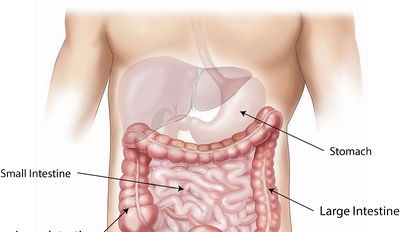 The main function of the stomach is chemical and mechanical processing of food. The first is carried out mainly by enzymes of gastric juice, which break down food substances (mainly proteins) and prepare them for absorption. Mechanical processing of food, that is, grinding it, mixing with gastric juice and moving from the stomach into the intestine, is carried out due to peristalsis (muscle contractions) of the stomach. The main function of the stomach is chemical and mechanical processing of food. The first is carried out mainly by enzymes of gastric juice, which break down food substances (mainly proteins) and prepare them for absorption. Mechanical processing of food, that is, grinding it, mixing with gastric juice and moving from the stomach into the intestine, is carried out due to peristalsis (muscle contractions) of the stomach.
The activity of the stomach as one of the main parts of the digestive system was especially thoroughly studied by the great Russian physiologist I.P. Pavlov and his students. The basic laws of gastric digestion were revealed and the leading role of the nervous system in the regulation of stomach activity was established.
IP Pavlov in the process of digestion identified two phases: conditioned reflex and neuro-humoral. The reflex phase coincides with the act of eating, when the secretion of gastric juice occurs under the influence of neuropsychic influences - the smell of food, its type, table setting. These influences are transmitted through the sensory organs to the cerebral cortex, and in response, even before eating, there is an abundant secretion of gastric juice, which Pavlov called "fiery".The release of juice continues after, when eating under the influence of gustatory sensations, the act of chewing and swallowing. In this second phase of digestion, secretion of juice is mainly supported by chemical pathogens contained in food, which are absorbed into the bloodstream from the gastrointestinal tract. The secretion of the hormone secretin, which enhances the secretion of juice, also affects the secretion of juice.
IP Pavlov found that fats inhibit the secretion of juice in the stomach; boiled vegetables, bread, fruits, potatoes, meat and meat soup (broth), on the contrary, enhance. He also proved that with a long-term lack of table salt, juice secretion decreases until it stops completely.
The stomach is completely emptied from the food taken after 2-6 hours, depending on its quality. Meat and fats are retained in the stomach for the longest time, water and milk leave it the fastest. Fat causes a strong contraction of the pylorus, and this delays the passage of food into the duodenum for a long time.
The intestine begins immediately behind the pylorus of the stomach and is a tortuous tube ending in the anus. It distinguishes between the duodenum, the small intestine, consisting of the jejunum and the ileum, and the large intestine.
The duodenum got its name because its length is equal to the diameter of 12 fingers, ie, approximately 23-27 centimeters. It is closely connected with the pancreas, has a horseshoe shape and consists of three sections: upper horizontal, descending and lower horizontal.
In the cavity of the duodenum enter: bile from the bile duct - and pancreatic enzymes, which are important in digestion.
The large intestine, about 1.5-2 meters long, is a continuation of the small intestine and is divided into six segments: the cecum with the appendix, the ascending colon, the transverse colon, the descending colon, the sigmoid colon and the rectum.
The small intestine has a length of about 6 meters, it is separated from the large intestine by a bauginia flap, which allows intestinal contents to pass only in the direction of the large intestine and prevents its return from the large intestine to the small intestine.
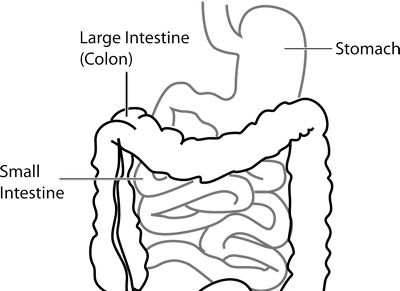 The cecum acquired its name due to its peculiar structure, resembling a blind bag; its length and width are usually the same size (6-8 centimeters). A blindly ending appendix (appendix) departs from the cecum, which is 7-9 centimeters long and 0.5-1 centimeters across. The cecum acquired its name due to its peculiar structure, resembling a blind bag; its length and width are usually the same size (6-8 centimeters). A blindly ending appendix (appendix) departs from the cecum, which is 7-9 centimeters long and 0.5-1 centimeters across.
After the cecum, there is an ascending intestine. Rising vertically, it forms a bend near the liver and passes into the transverse colon, which also forms a bend near the spleen and descends downward and partly forward.This segment is called the descending colon, which then passes into the sigmoid colon. It is located in the left half of the abdomen and has the shape of the Greek letter E (sigma), hence its name.
The rectum is the final section of the large intestine, ending in the anus.
The digestion of food and its assimilation by the body is mainly carried out in the small intestine. With the help of various enzymes of the small intestines, proteins are broken down to the stage of amino acids, fats into acids and glycerin, and carbohydrates to the stage of monosaccharides. These products of digestion are absorbed by the villi of the small intestine: amino acids, mineral salts, and water-soluble vitamins - directly into the blood, fats and fat-soluble vitamins - mainly into the lymphatic vessels.
In the large intestines, firstly, the entire mass of indigestible and indigestible parts of food passes: undigested plant fiber, tendons, cartilaginous tissues, etc., secondly, a small amount of nutrients that did not have time to be exposed to enzymes in the small intestines, and thirdly, almost all intestinal enzymes, as well as bile and bile acids.
In the large intestines (blind and ascending), further digestion and absorption of the digestible parts of food and fiber occurs with the participation of enzymes penetrating from the small intestines and bacterial flora, with the formation of gaseous products - methane, hydrogen, carbon dioxide and organic acids - lactic, butyric, oxalic ...
In the transverse colon and descending colon, water is absorbed and feces are formed. Therefore, the contents of the cecum and ascending intestine are liquid or semi-liquid, in the transverse colon - soft, and in the lower parts of the intestine acquires a thick consistency. Of the 4000 grams of the contents of the small intestines that have passed into the large intestines, about 150-200 grams of formed feces remain.
The movement of the food mass and its final digestion is entirely carried out by the intestines, which secretes food waste and gases that are unsuitable for nutrition. On average, the movement of the ingested food through the intestines takes from 24 to 48 hours, and approximately during this time, food waste enters the rectum.
The advancement of the food mass is formed as a result of several coordinated processes. First, the contents of the intestine move from the small intestine towards the large intestine and further to the anus due to longitudinal contractions of the intestine; secondly, contractions in the opposite direction and pendulum-like movements are observed, as a result of which the food gruel is mixed and soaked with digestive juices. (These muscle contractions are called peristalsis.) The complex processes associated with the movement of intestinal contents are carried out by the central and autonomic nervous systems, in particular by the nerve plexus located inside the intestinal wall.
A.G. Ghukasyan - Gastrointestinal Diseases
|
 Before proceeding with the presentation of preventive measures that prevent the development of diseases of the gastrointestinal tract, it is necessary to at least briefly dwell on its anatomical and physiological characteristics.
 The main function of the stomach is chemical and mechanical processing of food. The first is carried out mainly by enzymes of gastric juice, which break down food substances (mainly proteins) and prepare them for absorption. Mechanical processing of food, that is, grinding it, mixing with gastric juice and moving from the stomach into the intestine, is carried out due to peristalsis (muscle contractions) of the stomach.
 The cecum acquired its name due to its peculiar structure, resembling a blind bag; its length and width are usually the same size (6-8 centimeters). A blindly ending appendix (appendix) departs from the cecum, which is 7-9 centimeters long and 0.5-1 centimeters across.