What is a cage? |
|
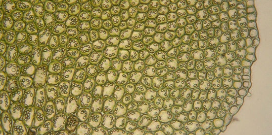
Two years later, he came across a cork. He made its thinnest cut and ... another discovery. His eyes saw the internal structure of the cork, resembling a honeycomb. He named these little cells "Cells", which in Russian translation means cells, nests, honeycombs, cells, in a word, something fenced off, isolated from the rest. This term was adopted by science, as it surprisingly accurately reflected the properties of elementary particles of living things. However, this became clear much later. In the meantime, different researchers are detecting cells in different objects. The idea of the universality of the structure of living matter is in the air. Biologist after biologist confirm: such and such a living organism consists of cells. The amount of observations is growing. A little more, and quantity should turn into quality. However, this "a little" took almost 100 years. Only in 1838-1839 the botanist Schleiden and the anatomist Schwann decided to generalize: "All living organisms are composed of cells." To say "all", science took more than a century, but this is the difference between the sum of observations and the scientific theory generalizing them. And yet, the cellular theory could not yet be considered created. The essential point remained unclear: where the cells themselves come from. Biologists have repeatedly observed and even described their division. But it never occurred to anyone that this process is the birth of new cells. One modern researcher rightly remarked in this regard: "Observation is rarely recognized if it forces us to draw unreasonable conclusions, and the statement that each cell arises as a result of the division of another, previously existing, seemed completely unreasonable."
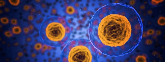
And yet, in 1859, an "unreasonable" postulate was formulated, which laid the foundation for a new cell biology: "Every cell is from a cell". The microscope of Robert Hooke was magnified 100 times. It was enough to see the cage. 300 years later, in 1963, an electron microscope enlarges a cell 100 thousand times. This is already enough to consider her. The difference, as physicists say, is only three orders of magnitude. But behind them is a complex and difficult path from descriptive biology to molecular biology, from the first acquaintance with the cell to a detailed study of its structures. The figure shows a cell as seen through a modern electron microscope. The reader should be patient: now her "inventory" will follow. We'll start with the shell. She is a cage custom. The shell vigilantly monitors that substances unnecessary at the moment do not penetrate into the cell; on the contrary, the substances that the cell needs can count on its maximum assistance. The nucleus is located approximately in the center of the cell. What it "floats" in is the cytoplasm, in other words, the contents of the cell. Unfortunately, there is little we can add to this far from exhaustive definition. We can’t even answer the most elementary questions unambiguously. Liquid cytoplasm or solid? Both liquid and solid. Does anything move in it or is everything in place? And it stands and moves. Is it transparent or opaque? Yes and no. What part of the cell does it occupy? From one percent to ninety nine. Everything is clear, isn't it? Nevertheless, the answers are correct. It's just that the cytoplasm is unusually changeable, it reacts to the slightest changes in the environment. Prick a single-cell amoeba with a needle, and you will see (of course, under a microscope) a lot of changes. The movement of the cytoplasm, its transparency, viscosity will change, the shape of the cell will change. In a word, act in any way on the cytoplasm, and you will see: it will definitely react somehow. In the cytoplasm, dissolved a huge amount of different? chemical substances. In it, many of them end their journey, and they often start at our table. We salt the soup - from it table salt gets into the cage. We put sugar in tea - it also reaches the cytoplasm, although on the way it breaks down in half into glucose and fructose. We eat fruits and vegetables - vitamins from them migrate into the cytoplasm. Finally, a cell always contains a large set of various proteins. All these substances do not stand idle, they work for the cell, in which it draws its strength, its future. However, the most surprising thing is not that these molecules have come together in the same place, but that they, albeit for a short time, coexist with each other. In a chemist's flask, many of these compounds and moments could not be held together - they would immediately react. But the cell is a wise politician, it needs to preserve the individuality of each molecule for its own purposes, and it takes every precaution.
So, the cytoplasm is the site of action of many chemical reactions taking place in the cell; in fact, it is the arena of its vital activity. But this arena is not an empty space; the living space of a cell is divided between its organs, or, as biologists say, organelles, which means the smallest organs. They divided among themselves not only the territory of the cytoplasm, they also clearly divided the spheres of influence. Organella number 1 - mitochondria, looks like a floating barge. If the mitochondrion is dissected, its internal structure resembles a narrow coastal strip of a sandy beach, on which waves have lathered up bizarre folds. Such folds of different thickness (in mitochondria they are called ridges) cross the entire inner space of the mitochondria. Mitochondria are the power stations of the cell. They accumulate energy, which then, as needed, will be spent on the needs of the body. These income and expense operations are carried out by the "main energetic" of the cell - adenosine triphosphoric acid, abbreviated as ATP. Moreover, it is interesting that both humans and bacteria store energy reserves in the same molecule - in ATP. When there is a need for energy - for a person, say, for muscular work, for mimosa - for rolling leaves, for fireflies - for glowing, and for a stingray - for the formation of an electric charge - requests come to the mitochondria, and thrifty dispatchers - special enzymes are split off from a large ATP molecule one or two pieces - a group of atoms containing phosphorus. At the moment of splitting off, energy is released. Electron microscopic photographs of cells taken several years ago clearly show the network stretching from the nucleus to the membrane - a whole collection of tubules, flagella, membranes, tubules. Even 30 years ago, when acquaintance with the cell could take place only through the mediation of a light microscope, no one really saw the network.Nevertheless, the scientists felt that there was "something" here, and persistently drew some cells in the cell. The electron microscope saw what the scientists had foreseen: it really turned out to be a network, and it was called endoplasmic, that is, intraplasmic. This network tightly surrounds the nucleus, mitochondria and organelles that are still unfamiliar to us - ribosomes. Ribosomes are protein cell factories. All living things are supplied with their products. Given the strategic importance of these facilities, nature has made sure that the work there was smoothly running. The productivity of the protein factory is enormous: per hour of operation, each ribosome synthesizes more protein than it weighs.
Chromosomes are found in the nuclei of all living things: bacteria, plants, animals. Human chromosomes look different from, say, a moth, but everywhere they serve the same service: they control protein synthesis. It is in the chromosomes that deoxyribonucleic acid molecules - DNA - are located. They, like a cookbook, contain recipes for preparing a huge variety of proteins that are used for the needs of the cell itself and for “export”. The normal functioning of the body is based on the strict specificity of tens of thousands of proteins. To keep your face in this commotion, you need to remember your own structure well. The squirrels themselves do not remember him; the cell does it for them with the help of DNA. One of its molecules stores the structure of dozens of proteins. Each chromosome is released a strictly defined amount of DNA for a given organism. The DNA in the chromosome is packed very tightly: the length of the chromosome is measured in thousandths of a millimeter, and the length of the DNA molecules placed in it is in meters. Now, when we consider a dormant, non-dividing cell, chromosomes are very poorly visible: they work, and for this they have to maximize their surface - they stretch and therefore narrow. However, this time does not last so long (for us) - only 10-20 hours. After a period of intense work, the cell begins to prepare for division; chromosomes are also preparing for it: they twist, thicken and line up all in one plane - at this moment it is easy to see them. By the time the reader comes to the description of cell division, the chromosomes will be clearly visible, and we, taking advantage of this, will tell about them in more detail. This is the end of our excursion into the cellular interior. But this does not mean at all that we have exhausted the cell; many of its details remained outside our attention. But we have chosen the main thing, without which it will be difficult to continue the path to our final goal. And, moving to it one more step, we need to take away from this chapter a clear idea of the three structures of the cell - the power station, the protein factory and the chromosome. If the reader got it, he got a pass to the next chapter. Azernikov V.Z. - The solved code Similar publications |
| Stepan Petrovich Krasheninnikov | Strength of the Earth |
|---|
New recipes
 In 1665, the Englishman Robert Hooke built a device that we call a microscope. Like any curious person, and scientists differ from a mere mortal among other advantages and this quality, Hooke began to examine everything that came to hand through a microscope.
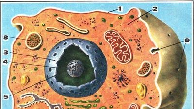
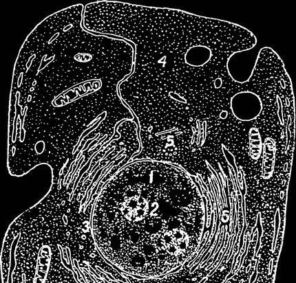 The modern scheme of the structure of the cell, based on electron microscopic observations: 1 - nucleus; 2 - nucleolus; 3 - nuclear envelope; 4 - cytoplasm; 5 - centrioles; 6 - endoplasmic reticulum; 7 - mitochondria; 8 - cell shell.
 To this end, it isolates some of the most aggressive molecules from their possible victims - it spreads the molecules in different "corners" of the cell - or, in extreme cases, humbles their chemical ardor. From the point of view of nature, this is done very ingeniously and simply (if one tried to implement the same technique in chemical laboratories, probably no one would dare to call it simple). What would each of us do if he needed to place a cat and a dog in the same room? Of course, I would muzzle the dog. Well, sometimes the cell does the same - it “puts on” enzymes - substances that govern all reactions in the cell, “restraining” molecules that close the active sites of enzymes.
 But like every business, ribosomes work under strict, unforgiving leadership. Orders come from the nucleus, from the main controller of protein synthesis - the chromosome.